
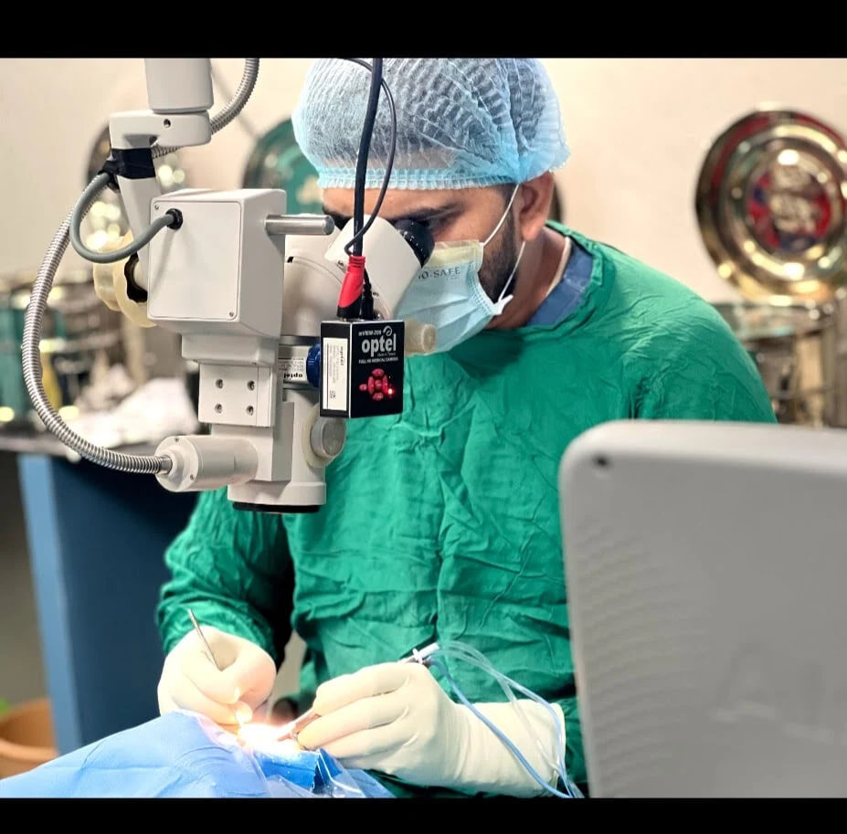
Eyelid trauma can damage the lid margin, tear canal, muscles or orbit. Repair aims to restore eye protection, drainage and a stable eyelid contour.
Understanding eyelid trauma
Cuts near the inner eyelid, full-thickness margin injuries and tissue loss require more than simple skin closure. Oculoplastic assessment considers both eye safety and appearance because the eyelids, orbit and tear drainage system work together. A treatment plan should therefore be based on the cause, severity, visual impact, ocular surface health, age, medical history and the patient’s priorities rather than on photographs alone.
For patients in Raipur and nearby areas, a consultation usually begins with a focused history and examination. Previous eye operations, thyroid disease, diabetes, blood-thinning medicines, trauma, contact-lens use and changes over time can all influence the diagnosis. Bringing old photographs, prescriptions and investigation reports can make the assessment more useful.
Common symptoms and signs
- Open eyelid cut or tissue loss
- Wound crossing the lash line
- Injury near the inner corner and tear drainage canal
- Difficulty opening or closing the eye
- Double vision, numbness or sunken appearance after trauma
- Foreign body, severe swelling or reduced vision
Symptoms do not always match the visible severity. A mild-looking eyelid problem may cause significant irritation or visual-field difficulty, while a dramatic cosmetic change may still require tests before treatment. One-sided, rapidly changing or painful symptoms deserve particular attention.
Why it happens
- Road traffic or workplace injuries
- Animal bites or sharp-object wounds
- Falls, sports injuries and assault
- Thermal or chemical burns
- Previous surgery or scar breakdown
- Associated facial or orbital fractures
Several conditions can look similar. For example, eyebrow descent can mimic an eyelid droop, dry eye can cause reflex watering, and thyroid eye disease can resemble a simple eyelid-position problem. Correctly identifying the main cause helps avoid ineffective or inappropriate treatment.
How an oculoplastic surgeon assesses it
- Immediate vision, pupil and globe-integrity check
- Careful wound exploration under appropriate anaesthesia
- Evaluation of the lid margin and canalicular system
- Eye-movement, sensation and orbital examination
- CT imaging when fracture or foreign body is suspected
Additional tests are selected only when they may change management. These can include visual-field testing, tear-drainage irrigation, clinical photography, blood tests, imaging of the orbit or sinuses, tissue biopsy, or review with another specialist. The exact work-up depends on the individual finding rather than a fixed package.
Treatment options
- Primary repair: Layered alignment of the eyelid margin and tissues when the wound is suitable.
- Canalicular repair: A silicone stent may support a torn tear canal near the inner corner.
- Tissue reconstruction: Local flaps, grafts or staged repair can replace missing skin and eyelid structure.
- Orbital repair: Fractures or foreign bodies may need separate treatment based on function and imaging.
- Scar revision: Later procedures can improve malposition or contour after tissues mature.
The safest option is not always surgery. Observation, lubrication, treatment of an underlying condition or a staged approach may be more appropriate. When an operation is recommended, the surgeon should explain the intended functional and cosmetic goals, anaesthesia, scars, realistic symmetry, possible need for revision, and condition-specific risks.
Preparing for treatment and recovery
- Tetanus and antibiotic decisions depend on the injury
- Keep wounds clean and avoid unapproved antiseptics near the eye
- Swelling can mask contour during early healing
- Follow-up checks eye closure, drainage, infection and scar contraction
Swelling and bruising are common after many eyelid and orbital procedures and often look worse during the first few days before improving. Cold compresses, head elevation, prescribed medicines and avoiding rubbing or strenuous activity are commonly advised, but instructions vary by procedure. Do not stop aspirin, anticoagulants or other prescribed medicines without approval from the clinician who manages them.
When to seek urgent care
Any eyelid injury with reduced vision, a misshapen pupil, severe pain, chemical exposure, suspected penetrating injury or uncontrolled bleeding is an emergency. Sudden loss of vision, severe eye pain, chemical injury, major trauma, rapidly increasing swelling, fever with worsening redness, or new double vision should be assessed urgently. Online information and contact forms are not substitutes for emergency care.
Questions worth asking at consultation
- What is the most likely diagnosis, and what alternatives need to be ruled out?
- Is the aim to protect the eye, improve function, improve appearance, or a combination?
- What result is realistic for my anatomy and medical history?
- What are the important risks, recovery milestones and warning symptoms?
- Will I need photographs, imaging, blood tests, biopsy or review by another specialist?
Key takeaway
Specialist repair aligns delicate eyelid structures and prioritises the eye itself before cosmetic refinement. A personalised examination is the best way to decide whether observation, medical treatment or surgery is suitable.
Frequently asked questions
Can any doctor stitch an eyelid cut?
Simple superficial cuts may be straightforward, but margin, inner-corner, deep or tissue-loss injuries benefit from specialist assessment.
Why is the inner corner important?
The canaliculi that drain tears run through the inner eyelids and may need stenting if cut.
When can scars be revised?
Many scars improve over months; revision timing depends on function, contraction and tissue maturation.
References & review notes
American Society of Ophthalmic Plastic and Reconstructive Surgery: https://www.asoprs.org/
NHS eye conditions and treatment information: https://www.nhs.uk/conditions/
Medical disclaimer: This article is for education only. Diagnosis and treatment depend on an in-person examination, medical history and, when needed, investigations.